Week 7
Nanoparticles present an interesting challenge when trying to confirm your product. They can't be confirmed in IR or NMR spectroscopy and are too small for visual confirmation. This means that the only way to really confirm the structure and presence of nanoparticles is the use of a transmission electron microscope.
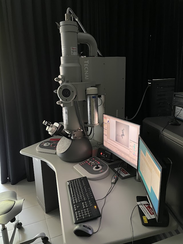
The TEM at the University of Fribourg. I'm not allowed to use it but I can take pictures.
TEM is one of several methods of electron microscopy. In electron microscopy, electrons are used to create images of objects in the electron beam. In TEM the image is created from the electrons that transmit (pass through) the objects. This allows us to get a view inside the objects we're trying to image. This is especially helpful for nanorattles, as we can get a peak inside.
There's loads of acronyms for different methods of electron microscopy; TEM, STEM, SEM, LEEM, PEEM, etc. SEM stands for Scanning electron microscopy. It creates an image based on the electrons that bounce off of the object being imaged. Here's an ant taken with SEM:

Personally I find electron microscopy fascinating. I hope you do too.


Comments
Post a Comment