Posts

Showing posts from July, 2023

Week 9

Image
My last post went over the basics of the nanoparticle synthesis that comprised about 1/3 of my time here. I figure that a good follow up to that post would be to talk about the organic synthesis that I did for the first 3 weeks of my time here. I've included the reaction scheme below as a visual aid.  The synthesis took a few days and the column purification was especially tedious, we ended up with just under a gram of the pure product.  Before coming here I was anxious that I wouldn't know what I was doing. I was pleasantly surprised when I realized that I understood the synthesis and was able to complete each step with minimal assistance.
Week 8 - Nanoparticle Synthesis

The past few days I've been working on my presentation for the end of this internship. I will end up giving it at least twice. Once to the research group here and once for the super-internship requirement. For today's post I'll be summarizing the synthesis of some of the particles I've been working with.  The synthesis begins with silver nitrate (AgNO3) (Blogger doesn't let me do subscripts, I'm sorry). It's combined with PVP-40 (a polymer) with ethylene glycol as a solvent. The mixture is then slowly brought to 120 degrees Celsius. The heat and PVP-40 allows the nanoparticle to form, with the PVP-40 acting as a stabilizing agent. The main (and only) method of purification in nanoparticle synthesis is centrifugation, as the particles are too small for filters, too light for columns, and cannot be allowed to dry.   This first step results in our silver nanoparticles. They are small, solid pieces of silver 30 to 50 nanometers in diameter. With the addition of

Week 7

Image
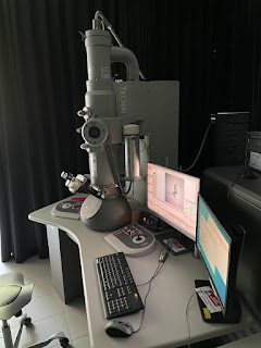
     Nanoparticles present an interesting challenge when trying to confirm your product. They can't be confirmed in IR or NMR spectroscopy and are too small for visual confirmation. This means that the only way to really confirm the structure and presence of nanoparticles is the use of a transmission electron microscope. The TEM at the University of Fribourg. I'm not allowed to use it but I can take pictures.        TEM is one of several methods of electron microscopy.  In electron microscopy, electrons are used to create images of objects in the electron beam. In TEM the image is created from the electrons that transmit (pass through) the objects. This allows us to get a view inside the objects we're trying to image. This is especially helpful for nanorattles, as we can get a peak inside. There's loads of acronyms for different methods of electron microscopy; TEM, STEM, SEM, LEEM, PEEM, etc. SEM stands for Scanning electron microscopy. It creates an image based on t